An echocardiogram – also known as an echo – is a non-invasive test that uses soundwaves to build up pictures of your heart. It’s the same kind of ultrasound technology that is used to scan pregnant women, as well as a range of other health applications.
Echocardiogram/ Echo
What is an echocardiogram?
How should I prepare for an echocardiogram? What happens on the day?
There is no specific preparation required. You can continue to take your medication as normal. However, it is recommended not to have caffeine for four hours prior to the scan.
Common questions:
- How long does it take? The duration of an echocardiogram varies from person to person and can range anywhere from 15 minutes up to an hour, but between 30 and 60 minutes is usual.
- What should I wear? You will be asked to remove the top half of your clothing and be given a gown to wear if necessary.
- Is it painful? It’s a very common, safe test, and most people find it’s not uncomfortable, though you may feel a bit of pressure as the sonographer applies the probe to obtain the best images. If you experience discomfort or pain at any point please tell the sonographer immediately.
- Can I bring someone with me? Our echocardiograms are undertaken by a trained cardiac sonographer in a dedicated, private clinic room. If you would like a chaperone, you can either bring someone with you or we can arrange one on your behalf.
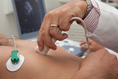
- What does it involve? Before your echocardiogram begins you will be asked to lie on a couch or bed. Three stickers will then be placed on your chest and arms to record the electrical activity of your heart. The sonographer will use specialist ultrasound gel to aid them in the examination (the gel may be cold and/or sticky but otherwise is completely harmless). Your sonographer will then take pictures from different locations on your chest and may ask you to change position so they can get the angles they need. It is normal for the sonographer to spend some time at each of these locations as there is a lot of information to be obtained. As the sonographer takes pictures you can expect to hear noises that represent the flow of blood through your heart. This is completely normal. The images they are collecting will allow your cardiologist to assess the structure and function of your heart. This includes examining the four chambers that make up the heart and the four heart valves that sit in between these chambers. The arteries (aorta/pulmonary artery) and veins (vena cava/pulmonary veins) that carry blood to and from the heart are also assessed.
On some occasions the sonographer may recommend that a small amount of contrast/dye is injected, to enable them to see the heart better. Alternatively, they may suggest that a small amount of bubbles are injected to check for any additional connections between the heart’s chambers.
Once the sonographer has all the images they need they will compile a report with all their findings.
We see both privately insured and self funding patients.
Please see our packages for self funding rates. We offer video consultations for patients who do not want to travel.
Learn about our packagesBook an appointment