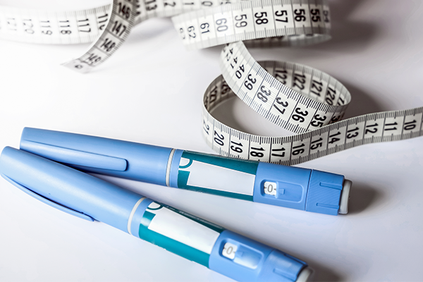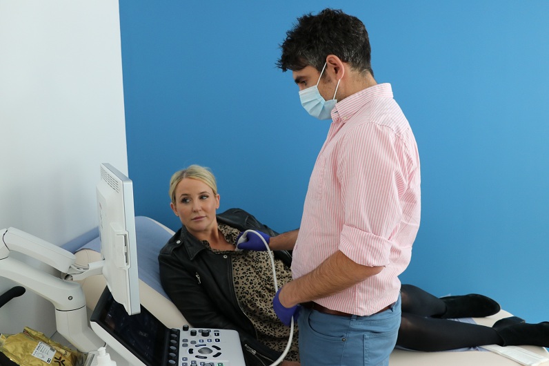
Echocardiograms are one of the most common heart tests in the world. Each year, they’re used to diagnose many cardiac abnormalities, which can range in severity.
These tests use a simple ultrasound to produce sound waves that create an image of your heart. An echocardiogram test at Venturi, Warrington (which is near Merseyside) can be used to diagnose a range of conditions and give your cardiologist a picture of your general heart health.
Having an echocardiogram test here at Venturi, Warrington is quick and non-invasive, and it’s one of the most reliable cardiac diagnostic tools in the world. Venturi has excellent access to the M62 and M6 making is very accessible to anyone in Manchester, Merseyside, Cheshire and throughout the North West.
So, what conditions can an echocardiogram diagnose? Let’s take a closer look.
Cardiovascular disease
Cardiovascular disease is an umbrella term used to describe a range of conditions that affect the heart. Some cardiovascular diseases are related to a process called atherosclerosis.
This condition occurs when a build-up of plaque develops around the walls of the arteries. Ultimately, this build up narrows your arteries and makes it harder for blood to flow freely through your body. If left untreated, this build-up can cause blood clots, heart attacks, or strokes.
The echocardiogram creates detailed images of your hearts valves and chambers. As a result, your doctor will be able to see the pumping action of your heart in real-time, which allows them to identify and diagnose conditions like atherosclerosis.
Atrial Fibrillation (AF)
Atrial Fibrillation is the most common type of heart arrhythmia, and it can interrupt the regular flow of blood through your heart. Thankfully, AF is treatable and rarely life-threatening. However, in serious cases, it can lead to blood clots in the heart.
In an episode of atrial fibrillation, the hearts upper chambers beat irregularly and out of sync with the lower chambers. An echocardiogram is the most simple and painless way to diagnose atrial fibrillation. Your doctor will be able to see where the heart muscle isn’t contracting the right way, which will help them identify the irregular rhythm and arrive at your diagnosis.
Pericardial effusion
Pericardial effusion is a condition that causes an extra build-up of fluid to collect between the heart and the pericardium (the sac that sits around your heart). This build-up puts additional pressure on your heart and may cause symptoms like shortness of breath, chest pain, or discomfort when breathing or lying down. The seriousness of the condition can depend on the primary cause and the size and rate of growth.
Echocardiography can be used to evaluate diseases in the pericardium, including effusion, constriction, and tamponade.
Heart valve disease
Heart valve disease can occur when one or more of your four heart valves do not work correctly. For example, heart valves may not open correctly, and some may not close tightly enough, causing blood to leak backwards across the valve (otherwise called ‘leaky valve’). Heart valve disease has several different causes, and in some cases, it can be life-threatening.
As echocardiograms build an image of your heart and its function, the ultrasound will allow your doctor to see your heart valves in action. They can then identify any abnormalities and determine which form of heart valve disease you may be suffering from.
Hypertrophic Cardiomyopathy (HCM)
Hypertrophic Cardiomyopathy can cause the heart muscle to become abnormally thick. This makes it much harder for your heart to pump blood around your body. Some people with HCM show no symptoms, while others may experience arrhythmias, fatigue, dizziness, chest pain, or shortness of breath.
An echocardiogram is an invaluable tool in the diagnosis of HCM. The ultra-sound technology can help doctors measure heart wall thickness and identify hypertrophy (an increase and growth in muscle cells) that occurs in unusual distribution.
Monitoring of heart conditions
In addition to being a rapid, painless, and accurate diagnostic tool, echocardiograms can also be used to monitor any pre-existing heart conditions. They allow cardiologists to carry out a systematic, in-depth study of cardiac structure and function, which can identify abnormalities and enable your doctor to assess a conditions response to medication and treatment. This makes it easier to monitor the severity of a condition, document its improvement, and alter treatment plans according to the results of your echocardiogram.
If you have any questions about any of the above, please do not hesitate to get in touch at hello@venturicardiology.com or on 01925 748 245.